We dare to say that we
may have just found a
secret weapon in the fight

against cancer. One that
explodes cancer cells.

“NOT YOUR GRANDFATHER'S HDM2”

Oncolyze is developing what we hope to be a transformational approach to cancer treatment. It all starts with a molecule called HDM2 (human double minute-binding protein-2). This is a protein that regulates cell growth. In fact, it counterbalances another protein called p53, which is a ‘tumor suppressor’. Most cancers overexpress HDM2 inside the cell, thereby blocking the p53 tumor suppressive function.

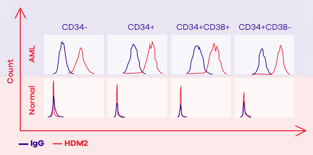
However, researchers at SUNY Downstate Medical Center (from where Oncolyze has licensed the technology) discovered that cancer cells have a high concentration of HDM2 not only intracellularly, but also on the external surface of their cell membrane. Whether this is used to somehow communicate with other cells, we are not yet certain, but what we do know is that more than 80% of cancer types we have tested have a high amount of HDM2 present on their cell surface, whereas normal cells appear to hardly have any. The unique expression of extracellular HDM2 on cancer cells makes it an excellent, selective target for an anti-cancer therapeutic agent.

Our therapeutic approach does exactly this. We target the extracellular HDM2 for a therapeutic mechanism that works at the cell surface and does not rely on the intracellular HDM2 and its function on p53. We know this because our drug candidates work on p53-null cell lines (cancer cells that lack p53 protein).

Our scientists and collaborators have discovered HDM2 on the surface of more than 20 types of solid and hematologic cancer cells.

In one example, HDM2 expression was shown to be dramatically different by flow cytometry in defined subpopulations (CD34-, CD34+, CD34+CD38+, CD34+CD38- cells) between cancer cells from leukemia (AML) patients and normal cells from healthy individuals.

HDM2 graph

KILLING CANCER CELLS VIA POP-TOSIS

drop

We have designed a series of fusion peptides that are able to target the surface-bound HDM2. A fusion peptide is a small protein that has two parts. Our lead drug candidate (OM-301) is a 32 amino acid peptide. The first 15 amino acids make up the HDM2 binding element; this sequence is derived from the HDM2-binding component of the p53 protein and is designed to bind HDM2 at the cell surface with high affinity. The remaining amino acids make up an amphipathic peptide, one that has both hydrophilic (water-liking) and hydrophobic / lipophilic (water-hating / fat-liking) elements.

When one end of OM-301 attaches to the cell surface HDM2, the other end wants to burrow its way into the cell membrane. Ideally, that part of OM-301 would probably prefer to go through the membrane and into the cell, but because the other end is attached to HDM2, it is forced to stay there. There is likely tension because some parts like to stay in the membrane, some parts like the extracellular water.

What happens next is outstanding. This tension causes pores or holes to be made in the cell membrane. The OM-301 almost acts as a ‘molecular icepick’. The overwhelming presence of holes causes the extracellular fluid to rapidly enter the cell via an osmotic mechanism. The cell rapidly enlarges, resulting in even greater tension on the stretched cell membrane, until it pops open. The cancer cell dies, not through the classical mechanism leveraged by most cancer therapeutics, known as apoptosis, rather a cell membrane mediated, biophysical mechanism for which we have coined the term ‘pop-tosis’.

cell
cell
cell
OM-301 peptides attach to the many HDM2 molecules on the surface of cancer cells.
Complexes of OM-301 + HDM2 form many small pores in the cell membrane of cancer cells.
Pores allow extracellular fluid to rapidly rush inwards, thereby causing the cell to swell and lyse.
Sample icon

Our peptides kill a variety
of cancer cells in vitro

Both OM-301 (our lead candidate) and OM-302 (our back-up candidate) have now been found to kill over 20 different cancer cell lines, including human melanoma, pancreatic, breast, colon, ovarian, lung, and ovarian cancers, primary ovarian cancer cells taken directly from patients, the human leukemia cell line K562, and primary acute myeloid leukemia (AML) leukemic and stem cells. When incubations of the cancer cells with either OM-301 or OM-302 were performed, the peptides killed cancer cells within minutes to hours.

In contrast, OM-301 and OM-302 have no or little effect on normal cell lines that have been studied, including pancreatic acinar cells, human fibroblasts, keratinocytes and normal breast epithelial cells. Importantly, they have no effect on the ability of human hematopoietic stem cells to differentiate in the presence of growth factors such as erythropoietin and colony-stimulating factor. This latter result, together with in vivo toxicity data, suggests that these peptides, unlike many chemotherapeutic agents, will have little effect on bone marrow; as a result, they are unlikely to cause anemia, pancytopenia and immune suppression.

OM-301 graphOM-301 graph
  • Normal Mouse Macrophages treated with OM- 301
  • K562 human CML p53 null cells treated with control
  • K562 human CML p53 null cells treated with OM-301

The in vitro effectiveness of OM-301 has been tested in about 20 cell lines, including p53 null cell lines, triple negative breast cancer, resistant human ovarian cancer & primary human AML stem cells.

The in vitro effectiveness of OM-301 has been tested in about 20 cell lines, including p53 null cell lines, triple negative breast cancer, resistant human ovarian cancer & primary human AML stem cells.

OM-301 is effective in
human tumors implanted into mice

Our collaborators around the world have shown efficacy of our peptides in various animal models of cancer. Most recently, our collaborators at City of Hope have shown effectiveness of OM-301 in a direct human transplant AML model, not only against ‘mature’ leukemia cells, but also the leukemia stem cells. Primary human AML CD34+ cells were transplanted into NSG-SGM3 mice that were subsequently treated with either OM-301 or vehicle for 2 weeks. The CD34+ cells were subsequently transplanted into a second group of mice and survival assessed.

OM-301 treatment resulted in significantly reduced self-renewal activity of human AML cells. At 4, 8, and 12 weeks, the % of CD45+ cells in the peripheral blood was significantly lower in the OM-301 treated mice. For example, by 12 weeks, the vehicle-treated mice had nearly 100% of CD45+ cells while those treated with OM-301 had approximately 60%. Furthermore, OM-301 almost doubled survival (p<0.0001). Only about 10% of secondary transplant mice survived at day 100, while all secondary transplant mice treated with OM-301 were alive at that point; no vehicle-treated mice survived past 110 days, while 50% of OM-301-treated mice survived past 150 days.

Although this was an initial proof of concept and not optimized for maximum therapeutic effect, it has presented us and our collaborators with undeniable promise for OM-301, especially in a difficult to treat cancer like AML.

Our collaborators at the City of Hope led by Dr. Guido Marcucci recently published an important scientific paper in the journal Leukemia with exciting in vivo preclinical data using OM-301 (previously, PNC-27) to selectively kill AML cells, including leukemic stem cells, with minimal off-target hematopoietic toxicity.

graph

Our lead
indication, AML

Acute myeloid leukemia (AML), our first indication, is the most common type of acute leukemia in adults. It is also called acute myelocytic leukemia, acute myelogenous leukemia, acute granulocytic leukemia, or acute non-lymphocytic leukemia. The American Cancer Society estimates about 20,000 new cases of AML in the US each year, but nearly 11,000 deaths from AML. Worldwide, it is estimated that there are about 1 million patients with AML, of which about 147,000 will die each year. AML is considered an orphan indication in the US, EU, and other geographies.

In AML, there is a rapid production of abnormal white blood cells that get collected in the bone marrow and disturb the production of normal blood cells. White blood cells guard the body against infections, and so patients with AML have a severely deteriorated immune system. AML is generally a disease of older people and is uncommon before the age of 45. The average age of people when they are first diagnosed with AML is about 68. As a result, these patients may have comorbid conditions that make treatment difficult or certain therapeutics intolerable.

About 50% to 75% of AML patients will achieve remission after initial therapy. Yet, even after complete remission, leukemic cells including leukemic stem cells, likely remain in numbers too small to be detected with current diagnostic techniques. Therefore, despite a number of recent advances in the treatment of AML, relapse remains common, the prognosis remains poor and treatment options for relapsed AML are quite limited. The only proven therapy is a hematopoietic stem cell transplant, if one has not already been performed, but it may not be available or possible for some patients. As a result, AML has a 5-year survival rate of only 25%, and long-term survival of almost zero.

PUBLICATIONS